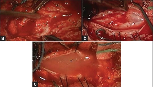
Figure 7.

(a) Scars and scattered pieces of the spinal cord as seen under surgical microscope at the beginning of surgery. (b) Reconstruction of the spinal cord. (c) At the end of surgery, PRP (biological scaffold) + stem cells in place, inside, and surrounds the damaged part of the spinal cord
