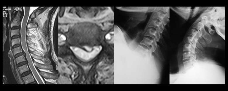
Figure 4.
Cervical spine magnetic resonance imaging (MRI) of a 54-year-old male patient who had suffered from neck and back discomfort for two years. This patient had symptoms that became more severe in the previous three months, with numbness of the limbs and unsteady gait. On examination, the patient had hyperreflexia of the patellar tendon, ankle clonus (+), bilateral Hoffmann’s reflex (finger flexor response) (+). The cervical MRI showed spinal cord compression in C5–C6 caused by left-sided cervical intervertebral disc herniation, with a high spinal cord signal at this level. The hyperflexion and hyperextension cervical spine X-ray showed C5–C6 segmental instability.

