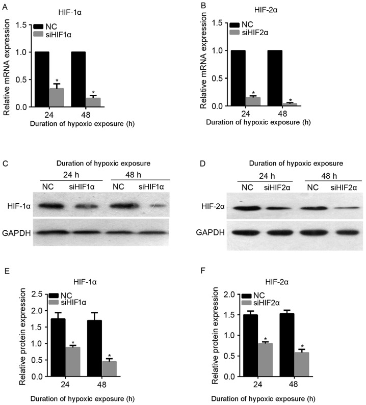
Figure 2.
mRNA and protein levels of HIF-1α and HIF-2α following siHIF1α and siHIF2α transfection under hypoxia. The mRNA levels of (A) HIF-1α and (B) HIF-2α following siHIF1α and siHIF2α transfection under hypoxia as detected by quantitative reverse transcription-polymerase chain reaction. The levels of protein expression of (C) HIF-1α and (D) HIF-2α following siHIF1α and siHIF2α transfection under hypoxia as detected by western blotting. Quantification of the expression levels of (E) HIF-1α and (F) HIF-2α in each group presented in bar graphs as fold increase. *P<0.05 vs. NC. HIF, hypoxia-inducible factor; NC, negative control; si, small interfering.

