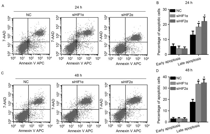
Figure 5.
Effect of HIF-1α and HIF-2α suppression on cell apoptosis in cervical cancer under hypoxic conditions. (A) Representative graph of flow cytometry results with Annexin V-APC/7-AAD staining of CaSki cells under hypoxic exposure for 24 h. (B) The percentage of early apoptotic and late apoptotic CaSki cells under hypoxic exposure for 24 h. (C) Representative graph of flow cytometry results with Annexin V-APC/7-AAD staining of CaSki cells under hypoxic exposure for 48 h. (D) The percentage of early apoptotic and late apoptotic CaSki cells under hypoxic exposure for 48 h. *P<0.05 vs. NC. APC, allophycocyanin; HIF, hypoxia-inducible factor; NC, negative control; si, small interfering; 7-AAD, 7-aminoactinomycin D.

