
Fig. 1.
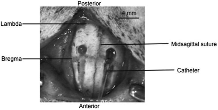
This picture shows the drilled troughs, with a catheter, made from PE-10 tubing, guided into place via the trough. The troughs are drilled 2–3 mm lateral of midline, and stop 1 mm posterior of bregma. The troughs are approximately 4 mm in length, gradually becoming deeper at the most posterior point. The skull is pierced and caution is taken not to penetrate the dura. Modified with permission from ref. 13.