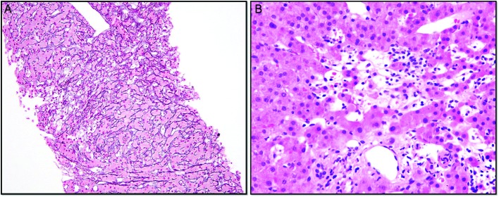
Figure 3.
(A) Core needle liver biopsy with reticulin and trichrome stains highlighting foci of hepatocyte dropout and collapse with areas of nodularity. Focal mild portal and sinusoidal fibrosis are present. (B) Core needle liver biopsy showing primarily lymphocytic inflammation with few eosinophils, neutrophils and macrophages, with an absence of basal plasmocytes.

