
Figure 4.
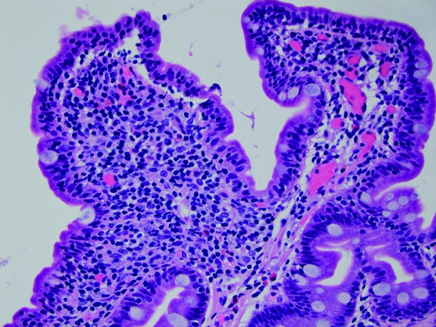
Sections demonstrate small intestinal mucosa with shortened villi with increased intraepithelial lymphocytes, a histological picture most commonly seen in coeliac disease but also seen with common variable immunodeficiency (H&E stain, 400× magnification). (Picture contributed by Dr Robert M Najarian, MD, Consultant in Gastrointestinal, Hepatobiliary and Pancreatic Pathology Central Pathologist, Pancreatic Disease Registry and Biorepository, Beth Israel Deaconess Medical Center, Department of Pathology, Boston, Massachusetts, USA.)