
Figure 3.
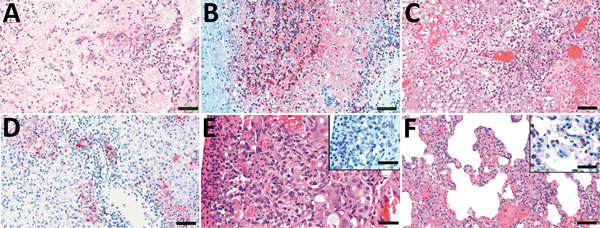
Histologic lesions and immunohistochemical detection of viral antigen in samples from ferrets exposed to live poultry market processing of highly pathogenic avian influenza A/Vietnam/1203/04 (H5N1) virus–infected chickens in study of airborne transmission of highly pathogenic influenza virus during processing of infected poultry. A) Olfactory bulb, 7 dpe, showing diffuse and severe neuropil malacia with mild cavitation and focal hemorrhages. Scale bar = 50 μm. B) Olfactory bulb, 7 dpe, showing viral antigen detected in neuropil, astrocytes, and neurons. Scale bar = 50 μm. C) Liver, 8 dpe, showing confluent coagulative necrosis of hepatocytes and bile duct necrosis with mononuclear cellular infiltrate in the portal triad. Scale bar = 50 μm. D) Liver, 8 dpe, showing viral antigen detected in hepatocytes, bile duct epithelia, and cellular debris. Scale bar = 50 μm. E) Nasal cavity, 7 dpe, showing moderate necrotic rhinitis with coagulative necrosis of mucous glandular epithelial cells; insert shows no viral antigen detected in mucosal membrane. Scale bars = 25 μm. F) Lung, 7 dpe, showing mild histiocytic interstitial pneumonia; insert shows viral antigen detected in type II pneumocytes. Scale bars = 25 μm. dpe, days postexposure.