Figure 1.

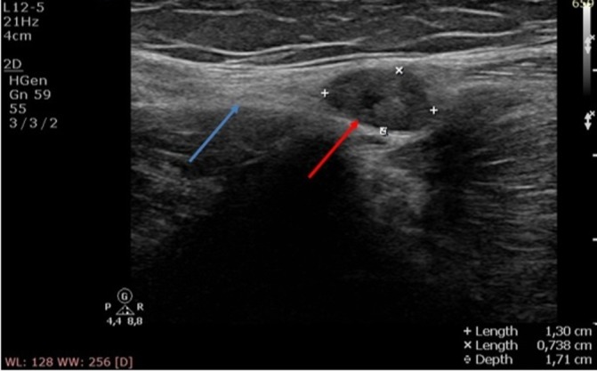
Ultrasound of the common peroneal nerve (blue arrow) demonstrating a 1.3 cm×0.74 cm echodense swelling behind the fibular head (red arrow).

Ultrasound of the common peroneal nerve (blue arrow) demonstrating a 1.3 cm×0.74 cm echodense swelling behind the fibular head (red arrow).