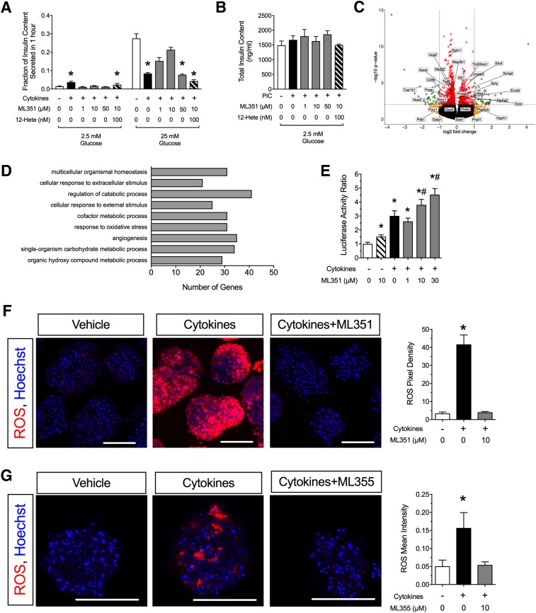
Figure 2.
12/15-LOX inhibition protects against cytokine-induced islet dysfunction and oxidative stress. A: Mouse islets were incubated for 24 h with cytokines, ML351, and 12-HETE before exposure to low (2.5 mmol/L) and high (25 mmol/L) glucose, after which insulin levels in the medium were measured (n = 3–4 independent experiments). *P < 0.05 compared with control conditions (no cytokines, no ML351, no 12-HETE). B: Total islet insulin content for the insulin release experiments shown in A. C: Volcano plot of RNA sequencing analysis of mouse islets treated with cytokines + 10 μmol/L ML351 vs. cytokines alone for 24 h (n = 3 independent experiments; red, genes significantly altered [P < 0.05]; yellow, genes altered more than twofold; green, genes significantly altered more than twofold). D: Gene ontology pathway analysis of data shown in C. E: βTC3 β-cells were transfected with Nrf2-Luc reporter and incubated with ML351 at the concentrations indicated and then treated for 6 h with cytokine before luciferase activity measurement (n = 10 independent experiments). Data are luciferase activity normalized to control conditions (no cytokines, no ML351). *P < 0.05 compared with untreated control (no cytokines, no ML351). #P < 0.05 compared with cytokine treatment alone (no ML351). F: Mouse islets stained for ROS (CellROX reagent) (red) and DAPI (blue) upon treatment with or without cytokines and 10 μmol/L ML351. G: Human islets stained for ROS (CellROX reagent) (red) and DAPI (blue) upon treatment with or without cytokines and 10 μmol/L ML355. Original magnification ×200; scale bars = 100 μm. Bar graphs show quantitation of CellROX intensity in 10–16 islets from three independent mouse islet preparations (or human donors). *P < 0.05 compared with untreated islets (no cytokines, no ML compound). Data are mean ± SEM. PIC, proinflammatory cytokines.

