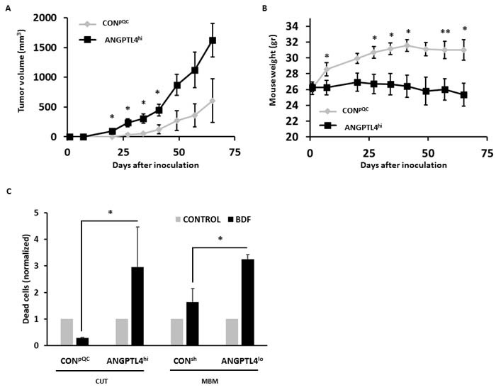
Figure 4. ANGPTL4 alters the tumorigenic potential of melanoma cells.

A. Volume of cutaneous tumors following subdermal implantation of CONpQC vs. ANGPTL4hi cutaneous cells. Tumor dimensions were measured using a caliper and volume was obtained as described in Materials and Methods. The average tumor volume + SEM is presented. *P < 0.05. B. Mice were weighed weekly following melanoma inoculation. The averege mouse weight + SD is presented. *P < 0.05. C. Melanoma cells were treated for 72 hrs with mouse BDF. Then, cells were trypsinized and cell death was determined by measuring DAPI incorporation. The bars represent the relative cell death of BDF treated cells compared to their controls + SEM obtained in one measurement in three independent experiments. *P < 0.05.
