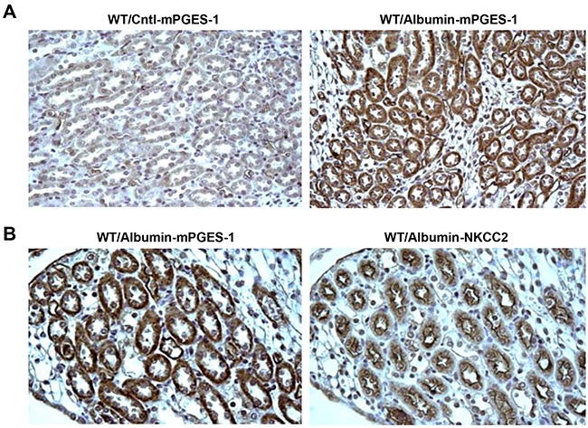
Figure 5. Localization of induced mPGES-1 in mouse kidney following albumin overload.

A. Immunohistochemistry of mPGES-1 illustrates the increase of mPGES-1 in tubules of albumin-loaded WT mice while consecutively stained section of kidney for albumin-loaded WT mice demonstrate B. mPGES-1 expression occurs specifically in the cells expressing NKCC2 (i.e. the TAL).
