
Figure 4. RECQ1 promotes DNA repair and survival after MMS treatment.
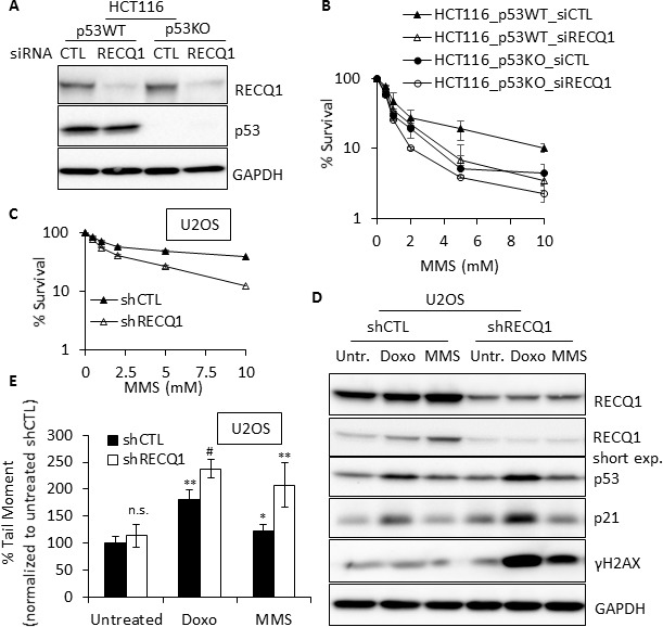
(A) Western blot showing siRNA knockdown of RECQ1 in p53-proficient and p53-deficient HCT116 cells. (B) RECQ1-depletion and p53 loss have synergistic effect on survival. Following 48 h of siRNA transfection, p53WT and p53KO-HCT116 cells were exposed to MMS for 24 h and subsequently grown for 24 h in drug-free medium. Surviving fraction compared to untreated was determined by cell count. Knockdown of RECQ1 was confirmed by Western blotting as shown. (C) U2OS cells stably transduced with a control (shCTL) or RECQ1 (shRECQ1)-specific shRNA were exposed to MMS for 24 h and subsequently grown for 24 h in drug-free medium. Surviving fraction compared to untreated was determined by cell count. (D) Western blot analysis of whole cell extracts of stable control and RECQ1 knockdown U2OS cells, untreated or treated with doxorubicin (1 μM) or MMS (1 mM) for 4 h. A short exposure of RECQ1 Western blot is also included. (E) DNA double strand breaks in control or RECQ1-depleted cells. Neutral Comet Assay was used to determine tail moment as a measure of double strand breaks in stable control and RECQ1 knockdown U2OS cells, untreated or treated with doxorubicin (1 μM) or MMS (1 mM) for 4 h. Mean tail moment of untreated shCTL was used to normalize the data and is shown as 100%. Statistical significance of difference in tail moment as compared to untreated shCTL is indicated as *p < 0.05; #p < 0.01; **p < 0.005 or n. s., non-significant.