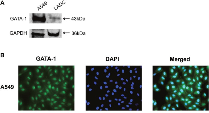
Figure 3. GATA-1 expression in human LADC tissue and A549 cells.

(A) GATA-1 protein expression was analyzed by immunoblotting in LADC tissue and A549 cells. (B) A549 cells were cultured for 24 h and analyzed by immunofluorescence. GATA-1 was immunostained in green and nuclei was stained with DAPI (blue).
