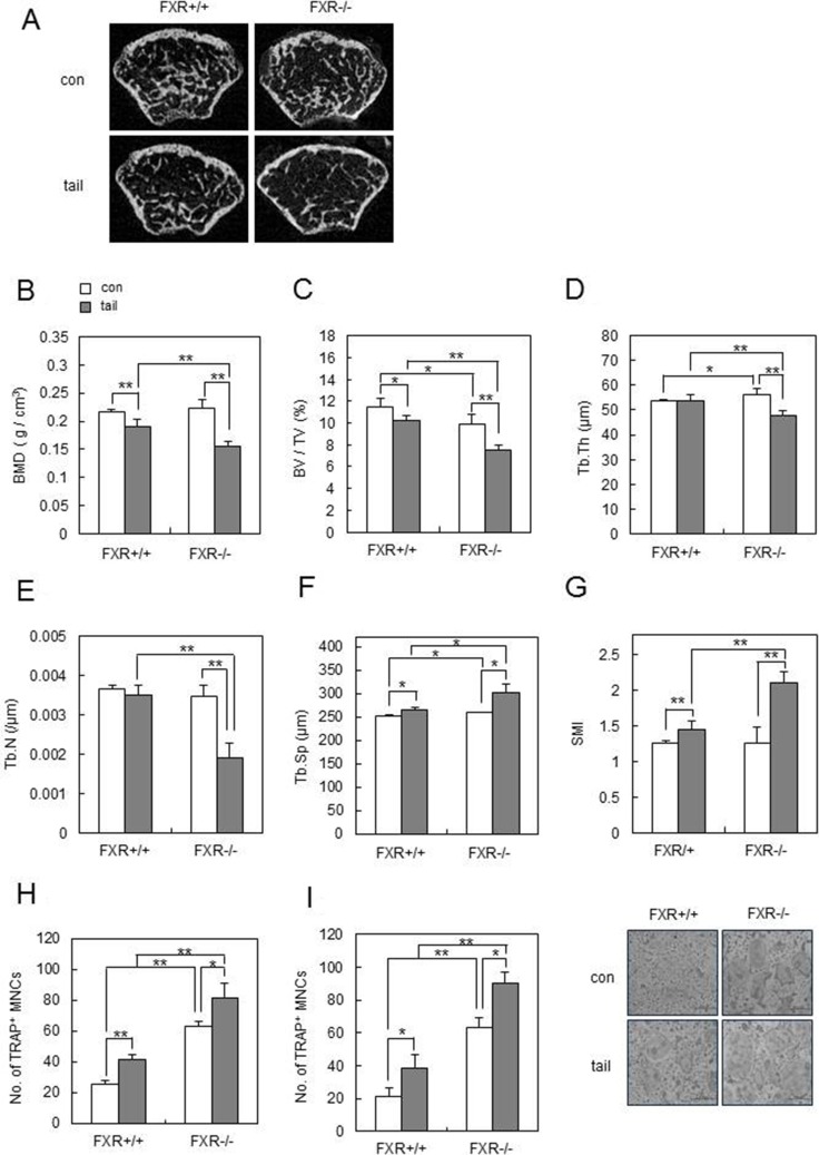
Figure 6. FXR deficiency accelerates unloading-induced bone loss in vivo.
Male 8-week-old FXR+/+ and FXR−/− mice were subjected to tail suspension, and mouse femurs were collected after 1 week. (A) Two-dimensional micro-CT images of the distal metaphysis of the femurs. (B–G) Various bone parameters of femurs were analyzed by micro-CT. BMD (g/cm3) (B), BV/TV (%) (C), Tb.Th (μm) (D), Tb.N (/μm) (E), Tb.Sp (μm) (F), SMI (G). (H–I) bone marrow cells (H) or BMMs (I) from FXR+/+ and FXR−/− mice were cultured with RANKL (100 ng/ml) and M-CSF (30 ng/ml) for 4 days and then TRAP+ osteoclasts were counted. *p < 0.05, **p < 0.01.

