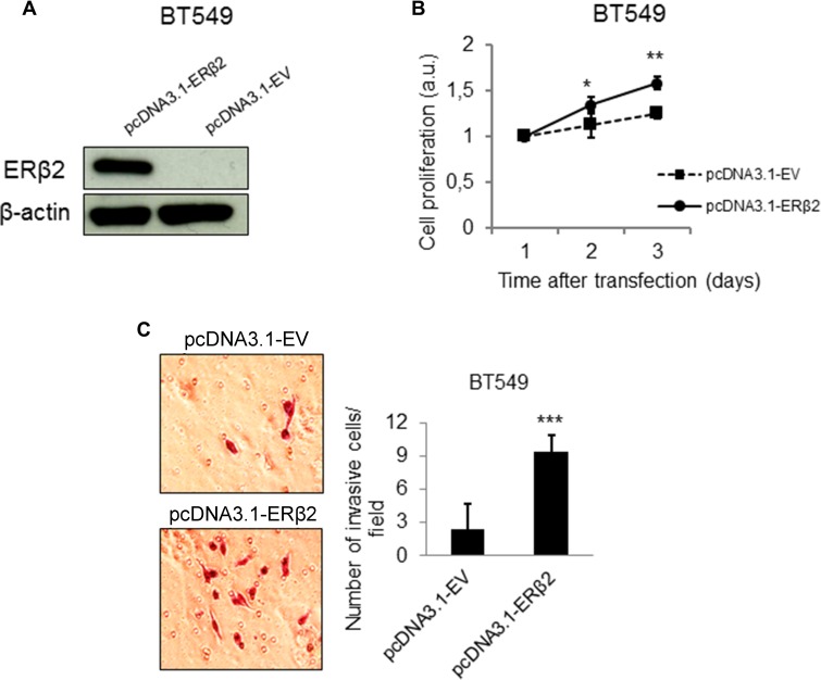
Figure 3. ERβ2 overexpression confers a more proliferative and invasive phenotype in vitro.
(A) Western blot analysis showing increased protein level of ERβ2 after transient overexpression of ERβ2 protein. ERβ2 was detected by the PPZ0506 antibody. β-actin was used as a loading control. (B) ERβ2 overexpression promotes cell proliferation in the BT549 cells. WST-1 assays of cell proliferation were carried out at the indicated time points after transfection of ERβ2 or empty vector (EV). Ratio of absorbance to day 1 is calculated. Data are shown as means of relative absorbance ± SD. *P < 0.05, **P < 0.01. Experiments were repeated three times. One representative experiment is shown. (C) ERβ2 overexpression promotes cell invasion in the BT549 cell line. BT549 cells were transfected with ERβ2 or EV, and cell invasion was evaluated by the BD Biocoat growth factor reduced Matrigel invasion chamber assay. Data represent means ± SD. ***P < 0.001. Experiment was repeated twice. One representative experiment is shown. B,C, p values were calculated by t-test.

