
Figure 1. Culturing tumor cells in NCS growth medium.
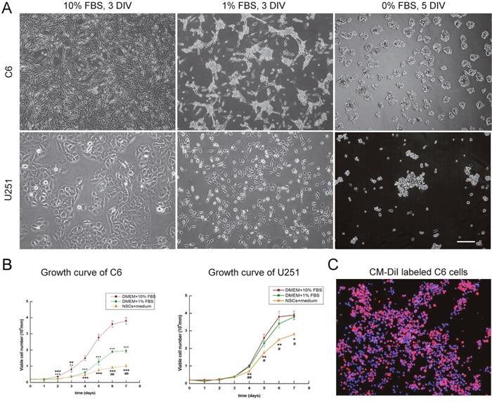
(A) C6 and U251 cells were cultured in DMEM with different concentrations of FBS. (B) Diverse growth of C6 and U251 cells in different mediums. (C) CM-DiI labeled C6 cells. DAPI used for cell nuclei staining showed in blue, CM-DiI was in red. Scale bars = 100 μm (A) and = 50 μm (C), DIV: days in vitro. *p <0.05.