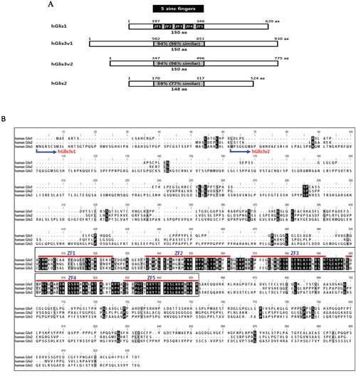
Figure 1. Sequence comparisons of the human Glis family proteins.

(A) The amino acid sequence homologies among hGlis proteins are shown in the 5 zinc finger-containing DNA-binding domains (grey box). The 5 black boxes in the DNA-binding domain of hGlis1 indicate zinc finger sequences. (B) The amino sequences of the hGlis proteins were aligned. Perfectly matched amino acids among hGlis1, 2 and 3 are represented by black boxes, and the amino acid positions with one mismatch among the hGlis proteins are marked in grey. The 5 red lines in the middle of the entire sequences indicate the 5 zinc finger sequences (ZF1-5). The N-terminal amino acids of the two variants of hGlis3 (hGlis3v1 and hGlis3v2) are marked at the beginning of the entire amino acid sequences.
