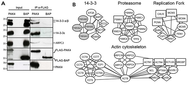
Figure 2. PAK4 interactome analysis reveals diverse cellular functions.

(A) Whole cell lysates derived from MCF7 cells stably expressing FLAG-PAK4 or FLAG-BAP were used for validation of QMS hits. After anti-FLAG IP and elution with FLAG peptides, samples were subjected to immunoblot analysis for the indicated proteins. Anti-FLAG (4th row) and anti-PAK4 (5th row) antibodies were used as controls. The left input panel shows immunoblotting of the two lysates. (B) PAK4 interactome networks obtained from the STRING database with the clusters identified by AutoAnnotate and visualized by Cytoscape. Diamond nodes: PAK4 interactors identified in the whole cell, or in both cytoplasmic and nuclear fractions or in all three fractions; Circle nodes: interactors identified in the cytoplasmic fraction or in both whole cell and cytoplasmic fraction; Squared nodes: interactors identified in the nuclear fraction or in both whole cell and nuclear fraction; Gray nodes: previously described PAK4 interactors.
