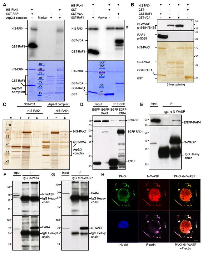
Figure 4. PAK4 interacts with and phosphorylates N-WASP.

(A) PAK4 mediated phosphorylation was analyzed by an in vitro kinase assay using recombinant HIS-PAK4 together with the Arp2/3 complex (left panel) or GST-VCA (right panel) as substrates, with GST as a negative control and GST-RAF1 (332–344) as a positive control (upper panels). The lower panels display the protein loading in the assays by Coomassie Brilliant Blue staining. (B) HIS-PAK4 phosphorylation of the WASP VCA domain was analyzed using an anti-N-WASP pSer484/Ser485 antibody after a kinase assay using recombinant HIS-PAK4 with GST-VCA as a substrate. GST serves as a negative control, while the anti-RAF1 pSer338 antibody was used as a positive control to detect GST-RAF1 phosphorylated by PAK4 (upper panel). The lower panel shows the loading of HIS-PAK4 protein and GST-fusion proteins used in the assay by silver staining. (C) HIS-PAK4 was pulled-down in the presence of GST-VCA or the Arp2/3 complex with Ni-NTA agarose and input (I), supernatant (S) and pellet (P) analyzed by silver staining. (D) IP of EGFP control or EGFP-PAK4 transiently expressed in H1299 cells analyzed by immunoblotting using an anti-N-WASP antibody (upper panel right two lanes). The left two lanes show immunoblotting of the input lysates. Anti-GFP was used to control the expression and IP efficiency in the lower panel. (E) N-WASP was immunoprecipitated with an anti-N-WASP antibody from lysates of H1299 cells transiently expressing EGFP-PAK4 and samples were analyzed by immunoblot using an anti-GFP antibody with the lysate input to the left (upper panel). Anti-N-WASP was used to control the expression and IP efficiency in the lower panel. (F) PAK4 was immunoprecipitated with an anti-PAK4 antibody from lysates of MCF7 cells, with rabbit IgG as a control, samples were analyzed by immunoblot using an anti-N-WASP antibody with the lysate input to the left (upper panel). Anti-PAK4 blotting was used to control IP efficiency in the lower panel. (G) N-WASP was immunoprecipitated with an anti-N-WASP antibody from lysates of H1299 cells, with rabbit IgG as a control, samples were analyzed by immunoblot using an anti-PAK4 antibody with the lysate input to the left (upper panel). Anti-N-WASP blotting was used to control IP efficiency in the lower panel. (H) PAK4, N-WASP and F-actin co-localized in the cell periphery after re-plating. FLAG-PAK4 was labeled with an anti-FLAG mab (Green), N-WASP with an anti-N-WASP antibody (Red), F-actin with SiR-actin (Purple) and Nuclei with Hoechst (Blue), Scale bar: 10 μm.
