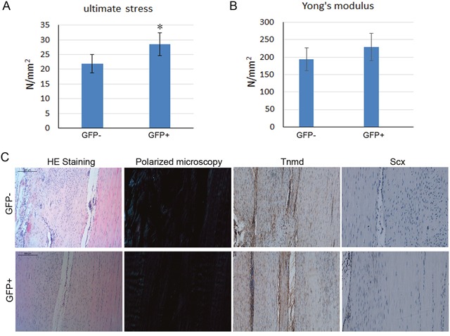
Figure 6. Effect of cell sheet formed by GFP positive and negative-rMSCs on tendon healing in rat patellar tendon injury model.

At 4 weeks after the operation, the tendon samples were collected for biomechanical testing and histological analysis. (A-B) The mechanical properties of the newly formed tendon tissues were analyzed by biomechanical testing. (C) The alignment of the collagen fibers was observed under polarizing microscope. The immunohistochemistry staining for Tnmd and Scx was also performed in the regenerated tendon tissue.
