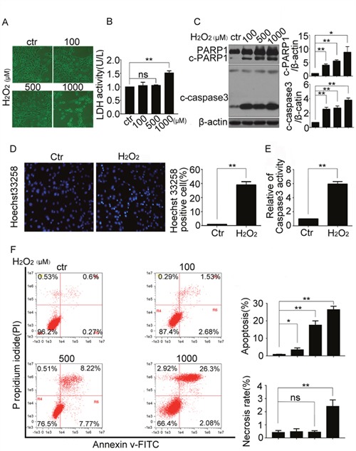
Figure 1. H2O2 induced apoptosis of resident cardiac stem cells.

CSCs were treated with indicated concentrations of H2O2 for 5 h. (A) The morphologic changes were observed with a phase contrast microscope (Scale bar=100 μm). (B) The LDH activity were detected by the LDH detection kit. (C) Western blot showed the protein levels of apoptosis-related proteins caspase3 and poly (ADP-ribose) polymerase 1 (PARP1). (D and E) CSCs were treated with H2O2 (500 μM) for 5 h, then the cells were stained with Hoechst 33258 to show the DNA fragmentation and condensation of CSCs (Scale bar=20 μm) (D), and caspase3 activity was analyzed (E). (F) The FCM showed the necrosis and apoptosis ratio of CSCs treated with indicated concentrations of H2O2. ctr, control. *P < 0.05; **P < 0.01; n=3.
