
Figure 1. Autophagic vacuoles are induced by DAC and DAS treatment.
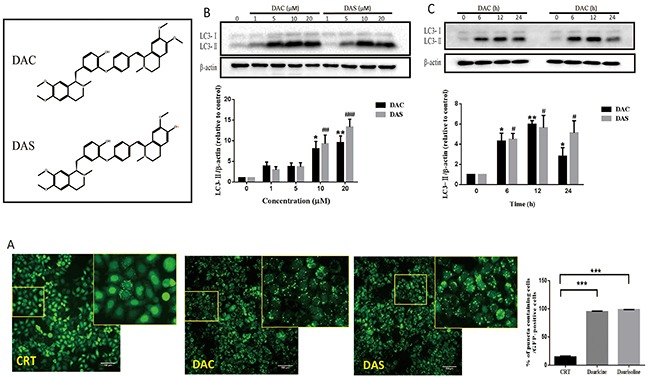
(A) GFP-LC3 puncta increase in DAC- and DAS-treated HeLa cells. The images of GFP-LC3-expressed HeLa cells were detected under InCell 2000 system after treatment of 10 μM DAC and DAS or vehicle (DMSO) for 24 h. Bars, 100μm. (B) DAC and DAS increased LC3 in a dose dependent manner. The LC3 expression in HeLa cells after treatment of different concentrations of DAC and DAS for 24 h; The relative intensity of LC3-IIexpression was calculated via Image J. (*P, #P < 0.05, **P < 0.01, ###P < 0.001 versus CRT). (C) LC3-II expression after treatment of DAC and DAS at different time points. The LC3 expression in HeLa cells at different time points after treatment with 10 μM DAC and DAS were analyzed via western blotting. The relative intensity of LC3-II expression was calculated via Image J. (*P, #P < 0.05, **P < 0.01 versus CRT). Error bars (mean±SEM). One way ANOVA with Turkey as post hoc tests.