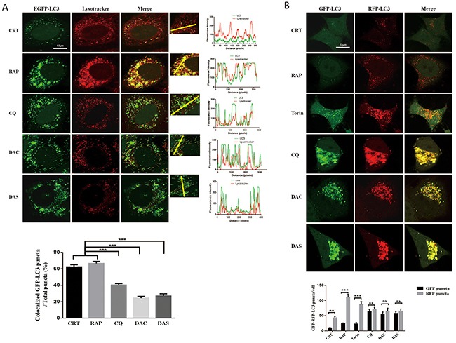
Figure 3. DAC and DAS inhibit the fusion of autophagosome and lysosome in HeLa cell.

(A) Co-localization analysis of EGFP-LC3 and Lysotracker. GFP-LC3 HeLa cells were treated with 10 μM DAC, 10 μM DAS, 30 μM CQ, 1 μM RAP, or DMSO for 24 h. The fluorescence images of LC3 and lysotracker were scanned with different channels via a confocal microscopy. The LC3 puncta that co-localizated with lysotracker were counted from at least 20 cells. Bar, 10μm. (B) Analysis of GFP-RFP-LC3 fluorescent signals in HeLa cells. HeLa cells were transiently transfected with GFP-RFP-LC3 plasmid, and treated with 10 μM DAC, 10 μM DAS, 30 μM CQ, 1 μM RAP, 200 nM Torin or DMSO for 24 h. The fluorescence images of LC3 were scanned with different channels under a confocal microscopy. Bar, 10μm. GFP or RFP puncta were counted at least in 20 cells.**P < 0.01, ***P < 0.001. Error bars are mean±SEM. One way ANOVA with Turkey as post hoc tests.
