Figure 1.
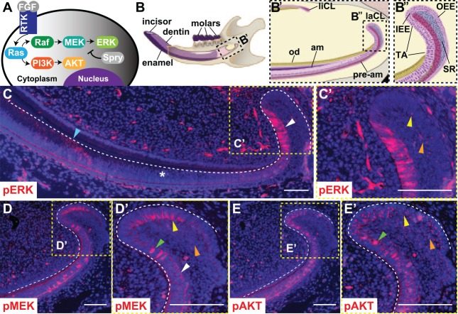
Phosphorylation of ERK, MEK, and AKT in the mouse laCL. (A) Schematic of the receptor tyrosine kinase (RTK)/Ras signaling pathway. Fibroblast growth factors bind to RTKs and activate Ras, which signals through Raf/MEK/ERK and PI3K/AKT. Spry proteins antagonize Ras signaling. (B) The mouse hemimandible contains 3 molars and 1 incisor, with the laCL and liCL present at the proximal end of the incisor. (B′) Magnified schematic of the boxed region in panel B shows the location of the laCL, liCL, pre-am, am, and od. (B′′) Magnified view of boxed region in panel B′ shows the different compartments composing the laCL: DESCs reside in the SR and OEE of the proximal laCL and give rise to the IEE and T-A cells. T-A cells generate pre-am, which differentiate into enamel-producing am. (C) Immunofluorescence with an antibody against pERK in the mouse incisor showed a biphasic expression profile with intense expression in T-A (white arrowhead), low or no expression in pre-am (*), and moderate expression in am (blue arrowhead) regions. (C′) Magnified boxed region shows scattered, low-intensity pERK staining in the OEE and SR (yellow and orange arrowheads, respectively). (D) pMEK was detected in the OEE, SR, and T-A. (D′) Magnified boxed region shows polarized membrane staining in the OEE (yellow arrowhead) and weak, evenly distributed membrane staining in the SR (orange arrowhead). In the T-A, pMEK staining was evenly distributed along the membrane (white arrowhead), or there was punctate nuclear staining (green arrowhead). (E) pAKT was detected in the OEE, SR, and T-A. (E′) Magnified boxed region shows weak, diffuse cytoplasmic staining and bright, punctate staining in the OEE and proximal SR (yellow and orange arrowheads). Cells in the T-A showed a similar pattern of diffuse and punctate cytoplasmic staining, plus nuclear pAKT staining (green arrowhead). Scale bars = 100 μm. am, ameloblasts; DESC, dental epithelial stem cell; ERK, extracellular signal-regulated kinases; IEE, inner enamel epithelium; laCL, labial cervical loop; liCL, lingual cervical loop; od, odontoblasts; OEE, outer enamel epithelium; pre-am, preameloblast; Spry, Sprouty; SR, stellate reticulum; T-A, transit amplifying.

