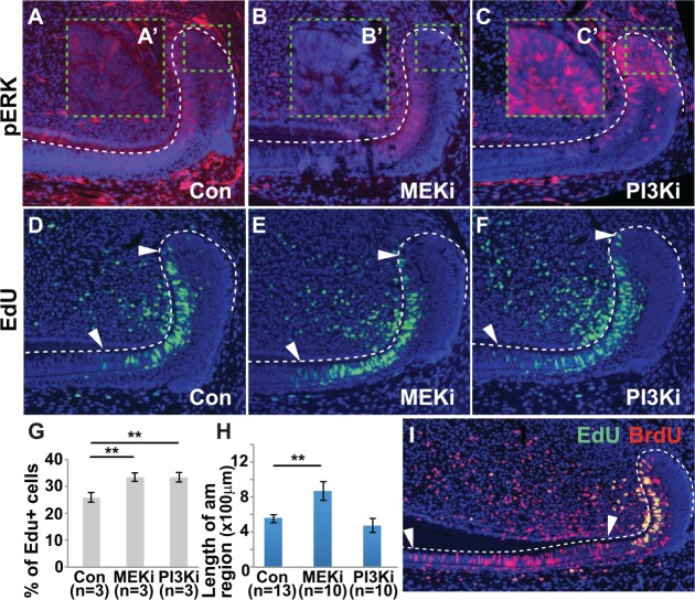
Figure 4.
Effect of MEK inhibition (MEKi) and PI3K inhibition (PI3Ki) on the labial cervical loop. (A–C) pERK staining in the labial cervical loop after oral administration of inhibitors for 7 d. (A′–C′) High-magnification images of the area of interest outlined by green dashed line in panels A–C. MEKi caused a decrease in ERK phosphorylation, particularly in the OEE and IEE (B, B′). Conversely, PI3Ki led to an increase in ERK phosphorylation, particularly in the OEE, IEE, and SR (C, C′). (D–F) Increased number of EdU+ cells was observed in the T-A region (between white arrowheads) with MEKi (E) or PI3Ki (F) as compared with control (D). (G) Quantification of the percentage of EdU+ cells in the T-A region. (H, I) Mice were coinjected with BrdU for 3 d and EdU for 2 h, and the length of the pre-am/am region was determined (between white arrowheads; I). The length of the pre-am/am region increased with MEKi treatment, but PI3Ki treatment did not affect pre-am/am length (H). am, ameloblast; IEE, inner enamel epithelium; OEE, outer enamel epithelium; pre-am, preameloblast; SR, stellate reticulum; T-A, transit-amplifying. Error bars represent SEM; ** P < 0.01.

