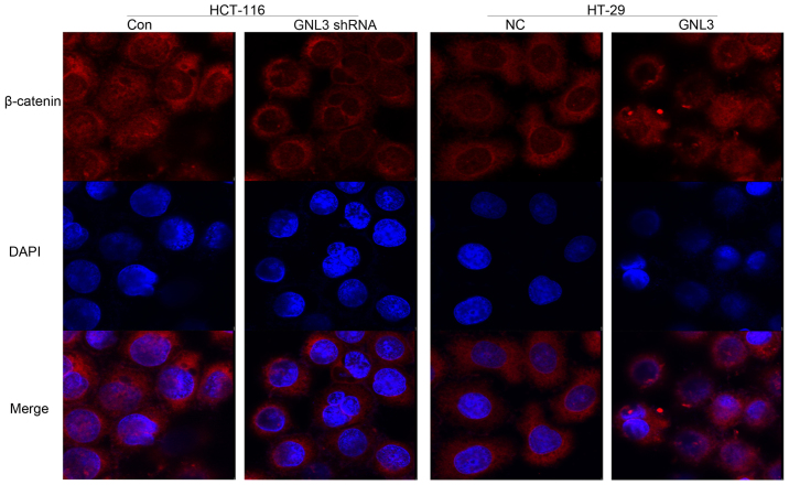
Figure 6.
Immunofluorescence staining for β-catenin. The β-catenin proteins labeled in red and the nuclei were stained with DAPI and are labeled in blue. GNL3 knockdown (GNL3 shRNA) in HCT-116 cells reduced the nuclear β-catenin levels compared with the controls (Con). However, GNL3 overexpression (GNL3) in HT-29 cells increased the nuclear β-catenin levels compared with the negative controls (NC). Original magnification, ×400.

