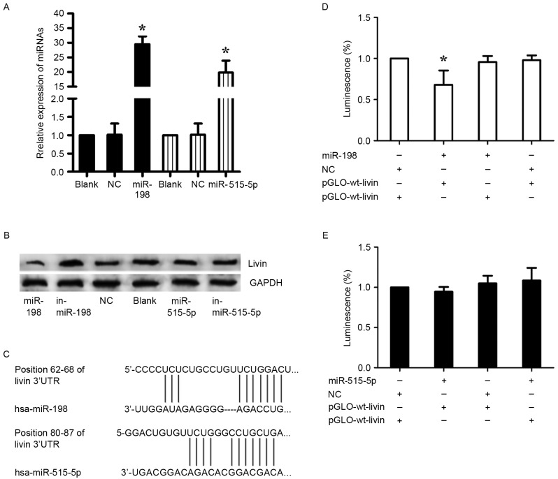
Figure 4.
Direct regulation of Livin by miR-198 in A549 cells. (A) The expression of miR-198 and miR-515-5p was evaluated by qRT-PCR in A549 cells 48 h after transfection to confirm the transfection efficiency. U6 was used as an internal control; *p<0.05. (B) The effects of miRNA mimics and inhibitors on Livin expression in A549 cells was detected using western blotting 48 h after transfection. GAPDH was used as a loading control. (C) The putative miR-198 and miR-515-5p binding sites in the 3′-UTR of Livin mRNA. (D and E) Dual-luciferase reporter assays using vectors encoding the putative miR-198 and miR-515-5p target sites in the Livin 3′-UTR for both the wild-type and mutant type. Normalized data were calculated as Renilla/firefly luciferase activity. In the miR-515-5p group, no significant change was observed. In the miR-198 group, the relative luciferase activity was clearly lower in cells cotransfected with the miR-198 mimics and pGLO-wt-Livin; *p<0.05 (the values for the pGLO-mut-Livin + NC group compared with pRL-TK vector were set equal to 1).

