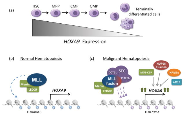
Figure 1.
Regulation of HOXA9 expression in normal and malignant hematopoiesis. (a) During normal hematopoiesis, HOXA9 is expressed most highly in early progenitor cells and its expression is subsequently down regulated as cells become terminally differentiated. (b) In normal hematopoiesis, HOXA9 expression is regulated by the MLL histone methytransferase, which deposits activating histone 3, lysine 4 trimethylation (H3K4me3) along the HOXA9 locus. This process requires interaction with menin and its cofactor LEDGF. (c) In approximately 50% of acute leukemias, HOXA9 is highly expressed as the result of a variety of upstream genetic alterations. These include MLL1-fusions, NUP98-fusions, MOZ-CBP fusions, NPM1c mutations and ASXL1 mutations. In the case of MLL1-fusions, one of 60 translocations partners is fused to the C-terminus of MLL, resulting in recruitment of the SEC (including DOT1L) and PTEF-b complexes. DOT1L is responsible for depositing activating histone 3 lysine 79 methylation, leading to high HOXA9 expression and malignant transformation. The other genetic abnormalities also result in high HOXA9 expression, though the mechanisms are less well understood.

