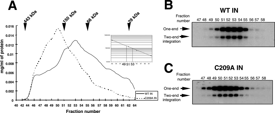
Fig. 7.
Chromatographic resolution of M-MuLV integrase proteins. (A) Elution profile of WT IN and C209A IN on S200 gel filtration chromatography. The relative position of migration of the globular molecular weight markers is indicated. The volume of each fraction was 275 µL. Inset: Migration is plotted vs the molecular weight of the markers. Dotted lines show molecular weight of fractions 50 and 53. The profile shows only fractions containing proteins. Aliquots (1 µL) of the fractions of WT IN (B) or C209A IN (C) were assayed in the concerted two-end integration assay, using a 28-mer 5′-labeled oligonucleotide donor substrate and target plasmid DNA as acceptor.

