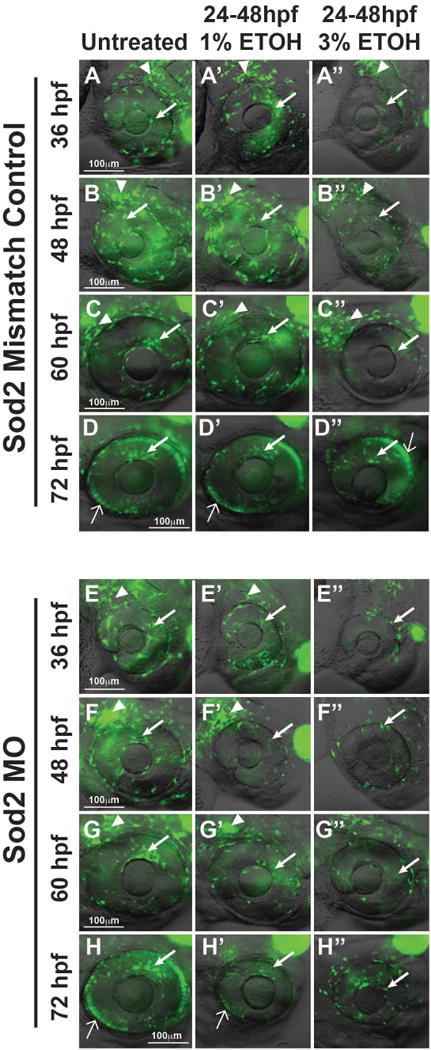
Fig. 7. Sod2 decreases effects of ETOH on ocular neural crest and anterior segment development.

Live imaging of Tg(foxd3:GFP) embryos showed that foxd3-positive cell migration into the anterior segment between 36 and 72 hpf was not affected by Sod2 MO knockdown (arrows, Fig. 7E–7H) compared to mismatch control-injected (Fig. 7A–7D) embryos. Knockdown of Sod2 in combination with 1% ETOH treatment from 24 to 48 hpf decreased foxd3-positive neural crest cell migration into the anterior segment (arrows, Fig. 7E′–7H′), but did not affect the cells in the periocular mesenchyme (arrowheads). This was in contrast to mismatch control-injected (Fig. 7A′–7D′) embryos in which 1% ETOH did not affect foxd3-positive cells in the periocular mesenchyme (arrowheads) or in the anterior segment migration (arrows). Treatment with 3% ETOH between 24 and 48 hpf decreased foxd3-positive cells in the periocular mesenchyme and in the developing eye in Sod2 knockdown (Fig. 7E″–7H″) and mismatch control-injected (Fig. 7A″–D″) embryos.
