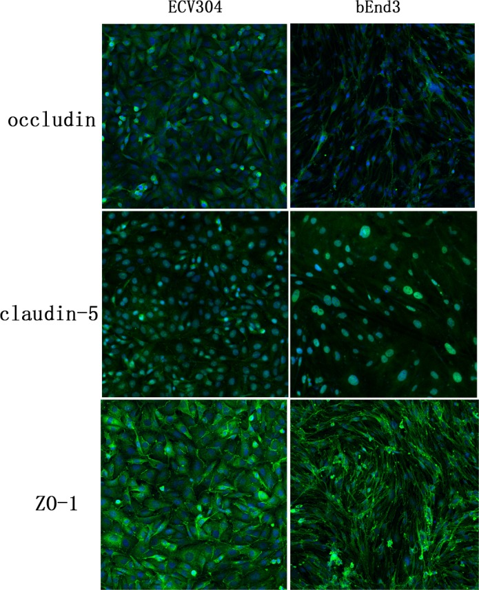
Fig 2. Immunofluorescent staining of tight junction proteins occludin, claudin-5 and ZO-1 in ECV304 and bEnd3 cells.

The immunofluorescence of ZO-1 gave distinct strands on cell membrane while the staining of occludin and claudin-5 were diffused and weak in both cell lines. The confocal images were acquired at 20 × magnification.
