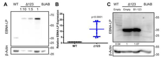
Figure 2.
The EBNA-LP protein accumulates to significantly higher levels in Δ123 LCLs. (A) Western blot of EBNA-LP expression in WT and Δ123 LCLs. The Δ123 LCL lysate was loaded at increasing concentration left to right, as indicated. BJAB lysate was loaded as a negative control. β-Actin was used as a loading control. (B) Δ123 LCL EBNA-LP expression normalized to WT LCL levels. Average of seven donors. p < 0.0001; one-sample t-test. Error bars = SD. (C) Western blot of EBNA-LP expression in WT LCLs transduced with the parental pTREX vector, Δ123 LCLs transduced with pTREX, and Δ123 LCLs transduced with pTREX expressing miR-BHRF1-123. BJAB cell lysate was used as a negative control. A long exposure is shown to allow the EBNA-LP level expressed in WT LCLs to be observed. EBNA-LP band intensity was first normalized to the loading control β-Actin and then to Δ123 LCLs transduced with pTREX.

