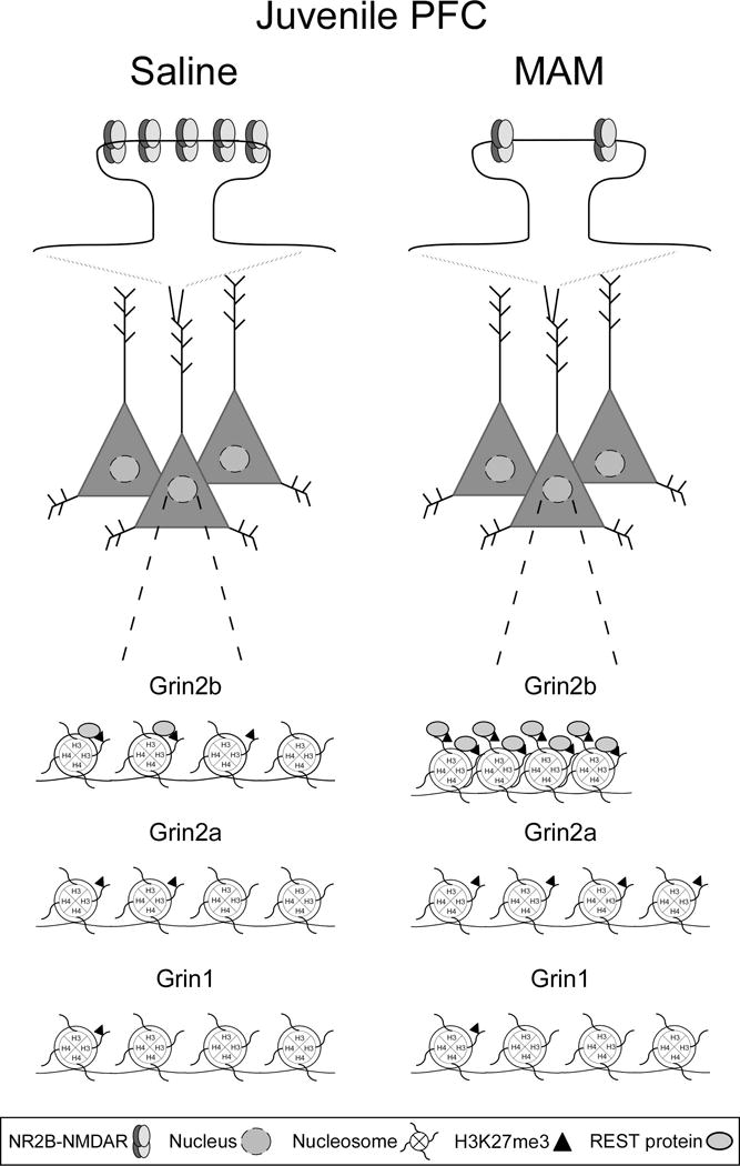
Figure 6.

A schematic representation of NR2B-NMDAR loss in the MAM juvenile mPFC, and REST (gray ovals) and H3K27me3 (black triangles) enrichment at response element (RE1) sites in the promoter regions of Grin2b, Grin2a, and Grin1 in juvenile saline and MAM mPFC. Top panel, NR2B-NMDAR levels are persistently high in the developing mPFC and play a critical role in synaptic plasticity and PFC-dependent cognitive functions. However, MAM animals are vulnerable to cognitive dysfunction due to the significant loss of synaptic NR2B-NMDARs in the critical juvenile period. Bottom panel, REST and H3K27me3 enrichment levels in saline mPFC are higher at the Grin2b RE1 site compared to the Grin1 and Grin2a RE1 sites, demonstrating selective REST regulation of Grin2b is an endogenous repressor mechanism. In the MAM mPFC, REST and H3K27me3 enrichment levels are greatly elevated compared to the saline group, demonstrating the hyper-repression of the proximal promoter region of Grin2b. Levels of REST and H3K27me3 enrichment are comparable between saline and MAM animals at the Grin1 and Grin2a RE1 sites, with the exception of a surprising elevation of H3K27me3 enrichment at the Grin2a promoter in MAM mPFC.
