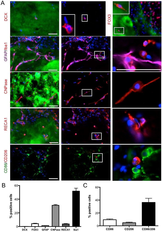
Figure 6. Phenotypic characterization of cells in ECM hydrogel.

A Immunohistochemical characterization of individual cell phenotypes within the ECM hydrogel at different magnifications. B. A phenotypic analysis of cells present within the hydrogel were predominantly microglia (ionized calcium binding adapter molecule 1, Iba-1), as well as fewer (p<0.01) cells from the oligodendrocyte lineage (2′,3′-cyclic-nucleotide 3′-phosphodiesterase, CNPase). Significantly (p<0.001) fewer neurons (Fox3), neural progenitors (doublecortin, DCX), astrocytes (glial fibrillary acid protein, GFAP) or endothelial cells (rat endothelial cell antigen 1) were found. C. A comparison of monocyte polarization indicated that these mostly co-expressed M1 (CD86) and M2 (CD206) markers, with significantly fewer cell expressing only CD86 (p<0.01) or CD206 (p<0.001). Scale bars 100 μm.
