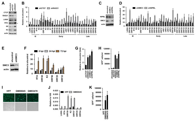
Figure 2. Cohesin factors are required for the restriction of lytic replication following KSHV infection.
(A) Immunoblot analysis of host and viral proteins in shCTCF- and shRAD21-treated SLK cells following KSHV infection. (B) Analysis of the effect of shCTCF and shRAD21 on viral gene expression at 72 hours KSHV postinfection. The classification of tested lytic viral genes based on their induction during KSHV lytic cycle is indicated such as IE (immediate early), early, and late. (C) Protein expression of the cohesin factors in shSMC3- and shNIPBL-treated SLK cells after KSHV infection. (D) Analysis of viral gene expression in shSMC3- and shNIPBL-treated cells at 72 hours KSHV postinfection using gene specific RT-qPCR. (E) Immunoblot showing the depletion of SMC3 expression in SLK cells upon shSMC3. (F) Time course RT-qPCR analysis of viral gene expression in shSMC3-treated SLK cells following KSHV infection. (G) The copy number of KSHV genome in different shRNA-treated SLK cells following KSHV infection was determined relative to shcontrol-treated cells at 72 hours KSHV postinfection. (H) KSHV titer was determined in the media of different shRNA-treated SLK cells at 72 hours KSHV postinfection. (I) Representative immunofluorescence images of KSHV BAC16-infected HFF and CdLS primary fibroblasts at 24 hpi. The KSHV clone BAC16 constitutively expresses GFP, which is used to detect infected cells. (J) RT-qPCR analysis of KSHV gene expression in infected HFF and CdLS fibroblasts at 24 hpi. (K) KSHV titer in the media collected from KSHV-infected HFF and CdLS fibroblasts at 72 hpi.

