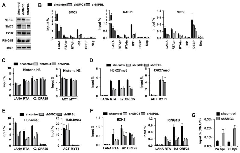
Figure 3. Effect of cohesin depletion on the deposition of histone marks onto the KSHV genome during de novo infection.
(A) Immunoblot showing the expression of host factors indicated on the left in lentiviral shRNA-treated SLK cells. (B) ChIP assay for SMC3, RAD21, and NIPBL on the indicated viral and host genomic sites in different cohesin factor depleted KSHV-infected SLK cells at 72 hpi. (C) Histone H3 ChIP, (D) H3K27me3 ChIP, (E) H3K4me3 ChIP as well as (F) EZH2 and RING1B ChIPs on viral and host genomic sites in shSMC3 and shNIPBL SLK cells at 72 hours KSHV postinfection. (G) RNAPII ChIP on RTA promoter in shcontrol- or shSMC3-treated SLK cells that were infected with KSHV for 24 or 72 hours. A two-tailed student’s t-test was applied to compare the H3 levels on the viral genomic regions between shcontrol and shSMC3 as well as between shcontrol and shNIPBL. For LANA, K2, and ORF25 p>0.05 while for RTA p<0.05.

