. 2017 Sep 28;58(6):1101–1110. doi: 10.3349/ymj.2017.58.6.1101

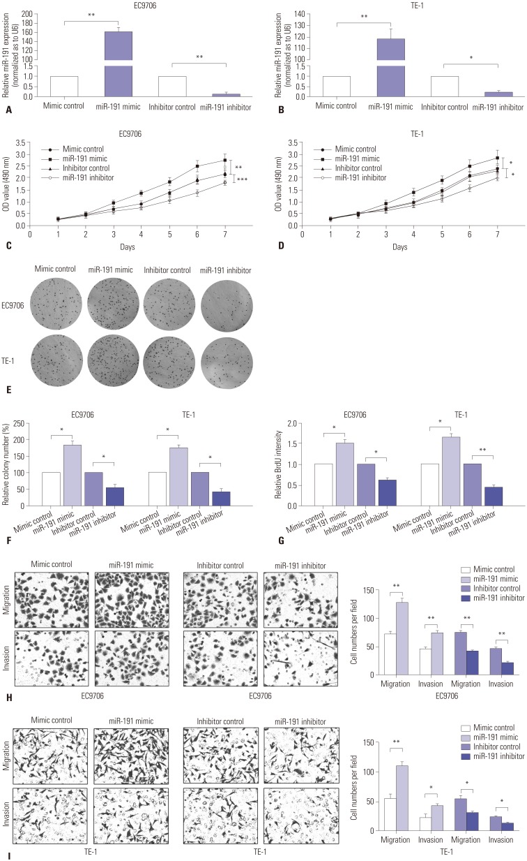
Fig. 3. miR-191 promotes ESCC cell proliferation and invasion in vitro. (A) RT-PCR analysis of miR-191 expression in EC9706 cells transfected with miR-191 mimic or inhibitor and corresponding control. (B) RT-PCR analysis of miR-191 expression in TE-1 cells transfected with miR-191 mimic or inhibitor and corresponding control. (C) MTT assay of EC9706 cells transfected with miR-191 mimic or inhibitor and corresponding control. (D) MTT assay of TE-1 cells transfected with miR-191 mimic or inhibitor and corresponding control. (E) The colony formation assays in EC9706 and TE-1 cells transfected with miR-191 mimic or inhibitor and corresponding control. Colonies were evaluated and values were reported as the ratio. (G) The BrdU assays from EC9706 and TE-1 cells transfected with miR-191 mimic or inhibitor and corresponding control. *p<0.05, **p<0.01, ***p<0.001 miR-191, microRNA-191; ESCC, esophageal squamous cell carcinoma; RT-PCR, real-time polymerase chain reaction; MTT, 3-(4,5-dimethyl-2-thiazolyl)-2,5-diphenyl-2-H-tetrazolium bromide; OD, optical density. (H) Transwell assays of EC9706 cells transfected with miR-191 mimic or inhibitor and corresponding control. Left panel: representative images. Right panel: quantification of 10 randomly selected fields. (I) Transwell assays of TE-1 cells transfected with miR-191 mimic or inhibitor and corresponding control. *p<0.05, **p<0.01. miR-191, microRNA-191; ESCC, esophageal squamous cell carcinoma.

Fig. 3