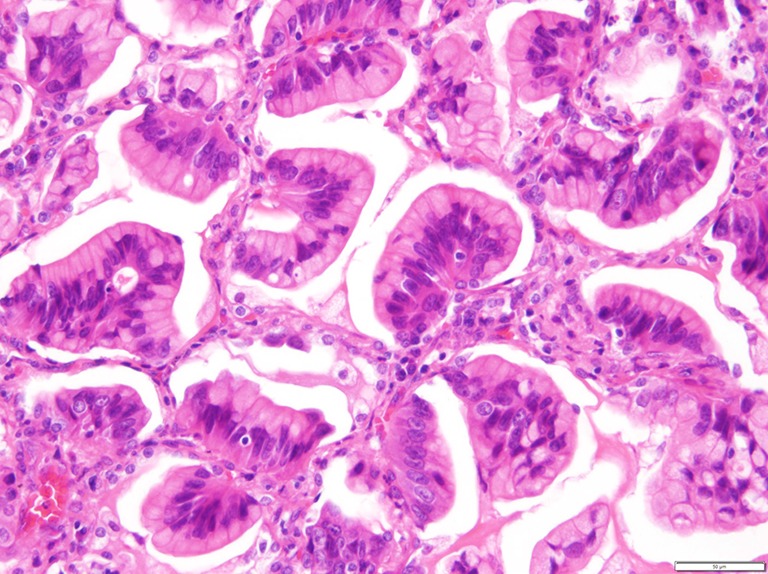
Figure 1.

A representative photograph of an invasive mucinous adenocarcinoma (IMA) demonstrating goblet or columnar tumor cells with abundant intracytoplasmic mucin and basally located nuclei, characteristic of IMA. (Hematoxylin and eosin stain, magnification ×200).
