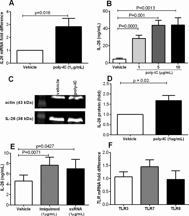
Figure 1.
Primary bronchial epithelial cells produce IL-26 enhanced by viral-related stimuli. Cells were stimulated (24 h) with different viral stimuli (TLR3 agonist poly-IC, TLR7 agonist imiquimod and TLR8 agonist ssRNA). Extracellular concentrations in cell-free conditioned media as well as intracellular expression of IL-26 protein were measured using ELISA and western blot, respectively, and levels of mRNA using real time. (A) IL26 mRNA levels after stimulation with poly-IC (n = 11). (B) Extracellular concentrations of IL-26 in cell-free conditioned media in response to poly-IC at different concentrations (n = 8). (C) Intracellular IL-26 protein (representative western blot). (D) the average protein expression (fold difference) after stimulation with poly-IC (1ug/mL) during 24 h. (E) Extracellular concentrations of IL-26 in cell-free conditioned media in response to imiquimod or ssRNA (n = 8). (F) TLR3, TLR7 and TLR8 mRNA levels (fold) (n = 5). The p values indicated are according to the Student paired t test. p < 0.05 is considered significant.

