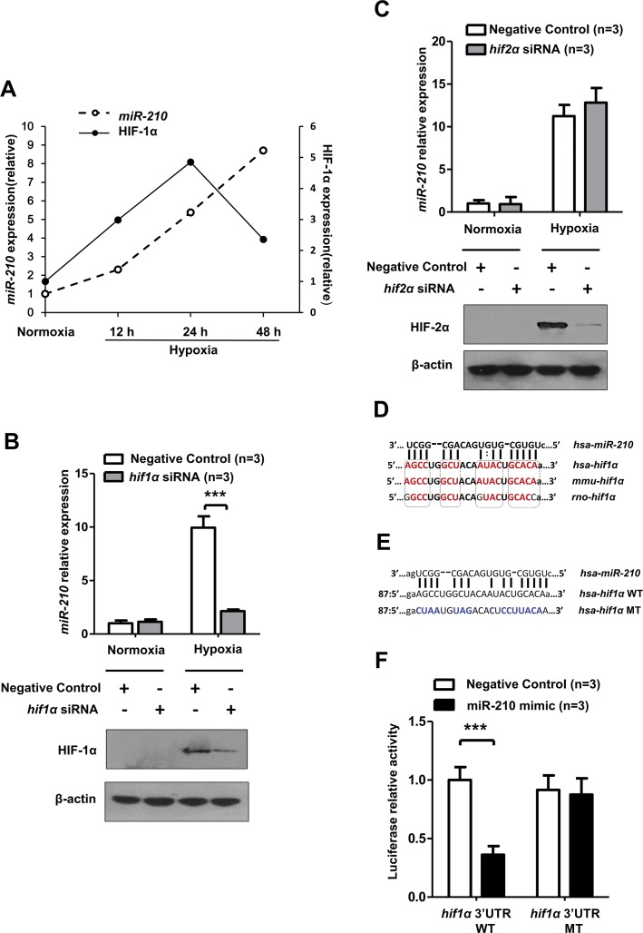
Figure 6.
miR-210 targeted 3′UTR of HIF-1α mRNA directly. (A) The relevance of miR-210 and HIF-1α protein expression in hypoxia-treated HK-2 cells (0.3% O2). The densitometry results for relative HIF-1α protein expression were normalized to β-actin. Detection of miR-210 expression after (B) HIF-2α and (C) HIF-1α siRNA transfection and in HK-2 cells. miR-210 expression was normalized to RNU6. The predicted binding site of miR-210-3p in the 3′UTR of human HIF-1α mRNA was found at the microRNA.org website (www.microRNA.org). (D) The potential binding site of miR-210-3p in the 3′UTR of HIF-1α mRNA is highly conserved among the selected mammals. The high conserved bases are shown in red. (E) The mutant-type (MT) design of the miR-210-3p binding sequence in the 3′UTR of human HIF-1α mRNA. WT, wild-type. (F) Luciferase reporter assays were conducted to determine the impact of miR-210-3p mimic on the activity of the 3′UTR of the human WT HIF-1α or the 3′UTR of the MT HIF-1α luciferase plasmid in HK-2 cells. Luciferase activity data were obtained after normalizing the firefly luciferase value to the renilla luciferase value. All quantitative results are from three independent experiments and are shown as mean ± SD. The Western blot data are representative results from three independent experiments. *P < 0.05; **P < 0.01; ***P < 0.001.

