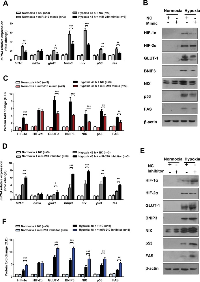
Figure 7.
miR-210 overexpression suppressed HIF-1α pathway activation in HK-2 cells after hypoxia, while miR-210 knockdown displayed the reverse effect. (A)Real-time PCR and (B) representative Western blotting were employed to detect the mRNA and protein expression levels of the HIF-1α pathway genes (HIF-1α, GLUT-1, BNIP3 and NIX), HIF-2α and the pivotal apoptotic genes (p53 and FAS) in HK-2 cells transfected with a miR-210 mimic and negative control under normoxia (21% O2) or hypoxia (0.3% O2) conditions for 48 h. (C) Optical density quantitative analysis of Western blots in (B). Regarding miR-210 inhibitor transfection, the mRNA expression levels of the abovementioned genes are shown in (D), and the corresponding protein expression are shown in (E). The optical density quantitative analysis of Western blots in (F). All quantitative results are from three independent experiments. All mRNA expression levels and Western blotting relative expressions were compared with β-actin (n = 3). Data are shown as mean ± SD. *P < 0.05; **P < 0.01; ***P < 0.001.

