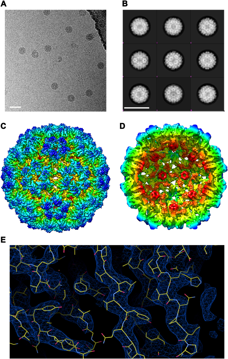
Fig. 2.
Structure resolution of engineered CMV VLPs by cryoEM. VLPs were dissolved in borate-EDTA containing buffer, vitrified and analyzed by EM using a Tecnai F30 ‘Polara’ microscope (FEI) a Representative 2-D view of vitrified VLP, generated from repeated imaging each field of view along the Z- axis creating a series of images (constituting a movie). From ~300 movies, 6685 particles were selected for data processing. b Examples of generated 2-D classification, 20 were generated based on average electron density. White bar corresponds to 50 nm. Cross sectional Selection refinement towards 3-D electron density model and an initial model fitting data to icosahedral symmetry produced 9 Å model. c High level refinement further improved resolution to 4.2 Å. In addition, d the internal view reveals ordered elements (shown in orange) are visible on the interior of the capsid shell. e Resolution Fitting reconstruction reveals that the obtained structure corresponds to X-ray crystal structural data

