Fig. 3.
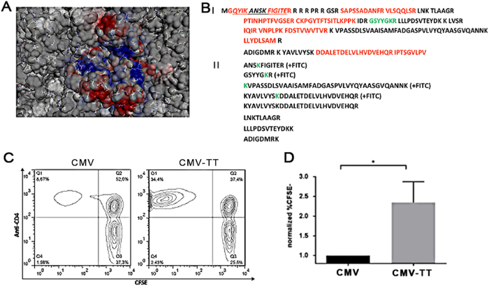
Chemical reactivity of surface exposed Lysine residues and T cell reactivity of incorporated universal T cell epitope. a Surface representation of cryoEM model with all lysine residues displayed in stick format, net surface charge is color coded. b Trypsin fingerprint analysis of CMV-Ntt830 VLPs after treatment with fluorescein isothiocyanate (FITC). Panel I—CMV-Ntt830 protein sequence represented in the form of trypsin peptides. Red—peptides identified after trypsin digest and mass spectrometric analysis; black—peptides not found; green—peptide identified only in the form of FITC conjugate. The Ntt830 residues are underlined; Panel II—CMV-Ntt830 coat protein peptides found as FITC conjugates (green Lys symbols) or partially cut by trypsin. c,d The universal T cell epitope in CMVTT is recognized by primary human CD4+ T cells. c Human PBMCs from four individual donors were labeled with CFSE and cultured for 7 days upon stimulation with either CMV or CMVTT and CFSE fluorescence was assessed by flow cytometry. Shown are representative scatterplots of CD4+ T cells stimulated with either CMV or CMVTT. d Normalized percentages of CFSE-CD4+ T cells in 4 individual donors upon CMV (black) or CMVTT (gray) are displayed as mean ± SEM. *p < 0.01 in a two-tailed, unpaired student’s t test

