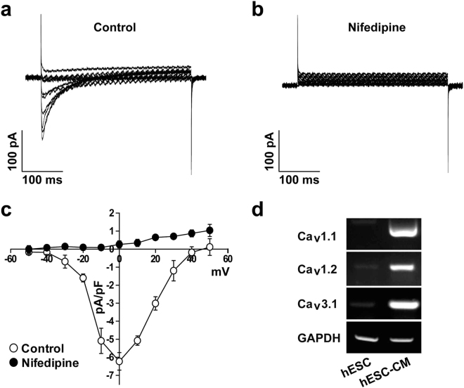
Figure 3.
Electrophysiological recordings of Ca2+ channels in hESC-CMs. Superimposed current traces were recorded by applying 500-ms step depolarizing pulses in the voltage range of −50 to +50 mV from a holding potential of −50 mV under control condition (a) and in the presence of 10 µM Nifedipine (b). (c) I-V relationship of peak current in the absence (open circle) and presence of Nifedipine (closed circle). n = 6. (d) Expression of Cav1.1, Cav1.2, and Cav3.1 subtypes in H9 cells and hESC-CMs was accessed by RT-PCR. n = 5. *P < 0.05.

