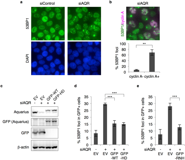
Figure 1.
R-loop-mediated DNA damage accumulates in Aquarius-depleted cells. (a) Representative images of 53BP1 foci formation in AQR-depleted cells. HCT116 cells were transfected with AQR or control siRNA, and immunostained 48 h later with anti-53BP1 antibody. (b) siAQR-induced 53BP1 foci formation in cyclin A-positive cells. At 48 h after AQR siRNA transfection, HCT116 cells were stained with anti-53BP1 and anti-cyclin A antibodies. The percentages of 53BP1 foci (>5)-positive cells in cyclin A-negative and -positive cells were determined. Data represent mean ± SD from three independent experiments. (c) Expression of GFP-tagged Aquarius (WT and HD). HCT116 cells were transfected with GFP-Aquarius (WT and HD) or the empty vector (EV) and then transfected with siRNA 24 h after plasmid transfection. Cells were lysed 48 h later with SDS sample buffer, and expression of the indicated proteins was analysed by western blotting. (d) Percentage of 53BP1 foci (>5)-positive cells in AQR-depleted and -complemented cells shown in (c). Data represent mean ± SD from three independent experiments. (e) Percentage of 53BP1 foci (>5)-positive cells in GFP-RNase H1-expressing siAQR cells. HCT116 cells were transfected with GFP-RNase H1 plasmid or the empty vector (EV), followed by siRNA transfection. At 48 h after siRNA transfection, cells were immunostained with anti-53BP1 antibody and the percentage of 53BP1 foci-positive cells was determined. Data represent mean ± SD from three independent experiments. **p < 0.01; ***p < 0.005.

