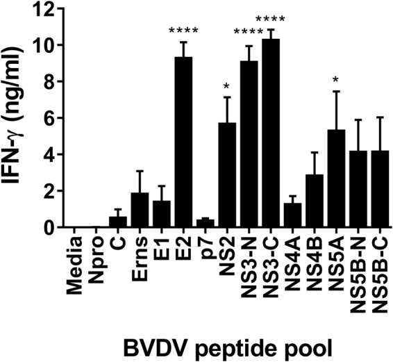
Figure 1.

Recognition of BVDV-1 proteins by T cells from immune cattle. PBMC from BVDV-immune cattle challenged by experimental infection with BVDV Oregon C24v (n = 5) were isolated on day 21 post-challenge, and stimulated in vitro with synthetic peptides pooled to represent BVDV-1 proteins. PBMC cultured in media alone were included as a negative control. IFN-γ secreted into culture supernatants was quantified by ELISA after 48 hours. Mean data are presented and error bars show the SEM. Values for virus and peptide pool-stimulated conditions were compared to the unstimulated (media) control using a one-way ANOVA followed by a Dunnett’s multiple comparison test; ****p < 0.0001, and *p < 0.05.
