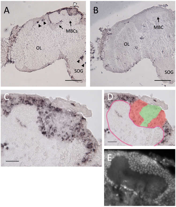
Figure 4.
In situ hybridization of Trp in parasitoid wasp A. reticulata adult female brain. (A,B) Frontal section of the brain hemisphere hybridized with antisense (A) and sense (B) Trp probes. Black arrowheads indicate Trp signals scattered outside MBs, and white arrowhead indicates the regions of Kenyon cells inside the calyx with a relatively weak Trp signal in (A). (C) Magnified view of the MB shown in (A). (D) Illustration indicating the Trp signal strength and the calyx is overlaid to (C). Regions with strong and weak Trp expression are colored with red and green, respectively. The calyx is outlined with red line. (E) DAPI staining of a serial section of (C,D). MBC, mushroom body calyx; OL, optic lobe; SOG, suboesophageal ganglion. Bars indicate 100 μm in (A) and (B), 20 μm in (C) and (D).

