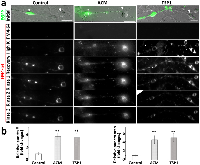
Figure 7.
The synaptic vesicle recycling study of connections between EGFP-ScNs and CN neurons. (a) EGFP-ScNs and CN neurons were co-cultured for 4–6 days, followed by FM4-64 synaptic vesicle recycling study. In initial exposure to FM4-64 for 1 min, no obvious staining was found in 3 groups. After high potassium stimulation, obvious staining was found in the soma and neurite outgrowths in the ACM and TSP1 groups. In the recovery stage, significant vesicle staining was observed in soma and nerve terminals of ACM and TSP1 groups. During rinse stages, the staining of vesicles remained distinct in the soma and neurite outgrowths. (b) The puncta at the recovery stage was used for the quantitative study. It was observed that relative number and area of FM4-64 vesicle along connections were lower in the control group, whereas they were significantly higher in ACM and TSP1 groups (mean ± standard error shown in the figure; **indicates P < 0.01, ANOVA followed by Tukey post hoc test). Scale bar: 25 µm in (a).

