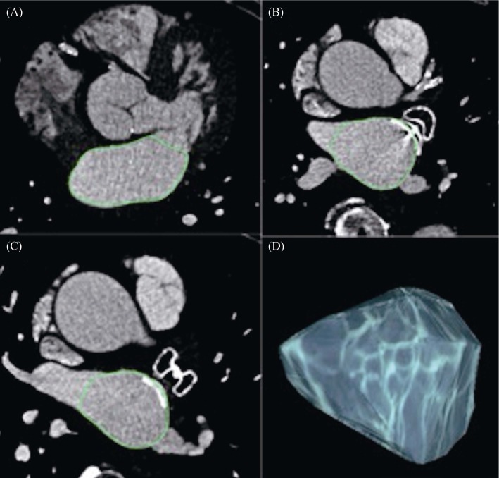
Figure 1. Measurement of left atrial volume using 3D volume threshold-based method of cardiac multidetector CT.
Volumetric assessment was performed on sets of axial images. The outline of the LA was traced manually on each slice, excluding the mitral valve (A) pulmonary veins (B, C) and the LAA (B, C). After selecting all the regions of interest within one series, OsiriX® automatically calculated the volume by multiplying surface and slice thickness and then adding up individual slice volumes. OsiriX® also provided 3D images using the “ROI volume” tool (D). CT: computed tomography; LAA: left atrial appendage; ROI: region of interest.

